Figure 1. In the Beginning
Biologists craft stories about the living world through practices of making, thinking, and communicating through images. The field of developmental biology in particular has retained, perhaps more than any other life science, its roots as a visual science in an increasingly data-infused era. Figure 1. In the Beginning draws inspiration from historical scientific figures and printed publishing formats to re-imagine what contemporary data presentation can look like. Using photogravure printing, it experiments with what happens when biological images are rendered in media now primarily considered the purview of art.
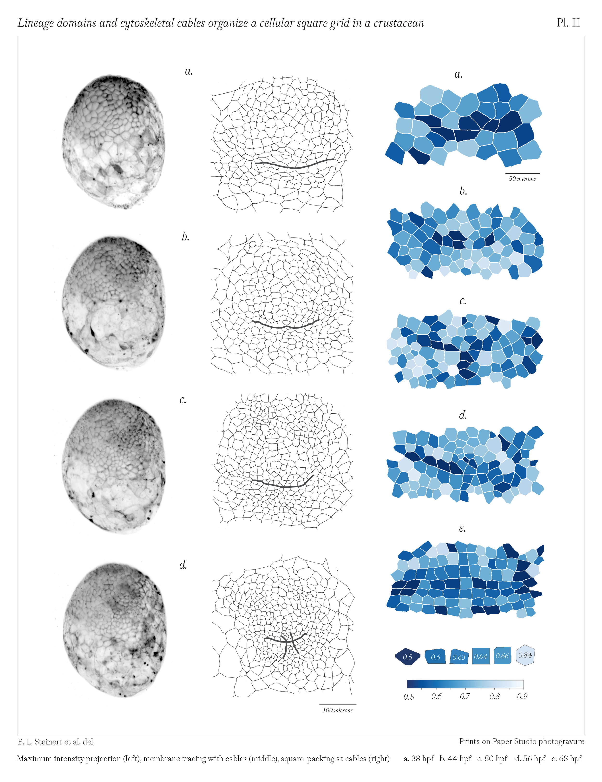
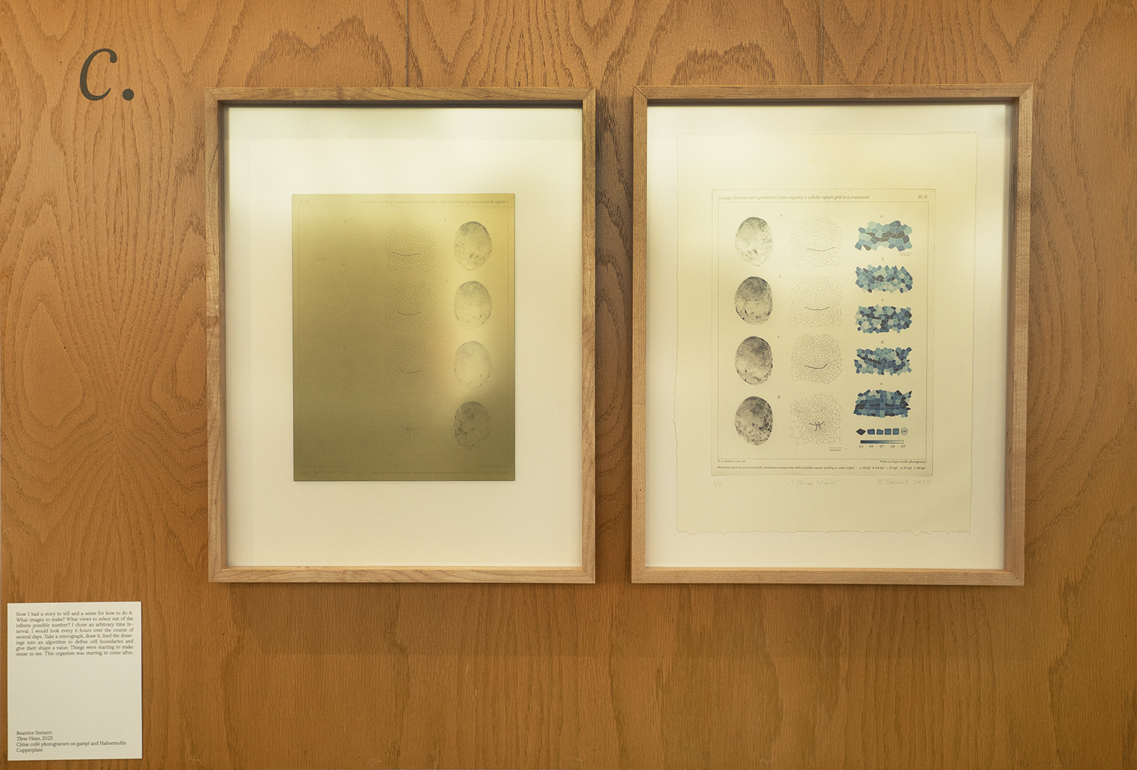
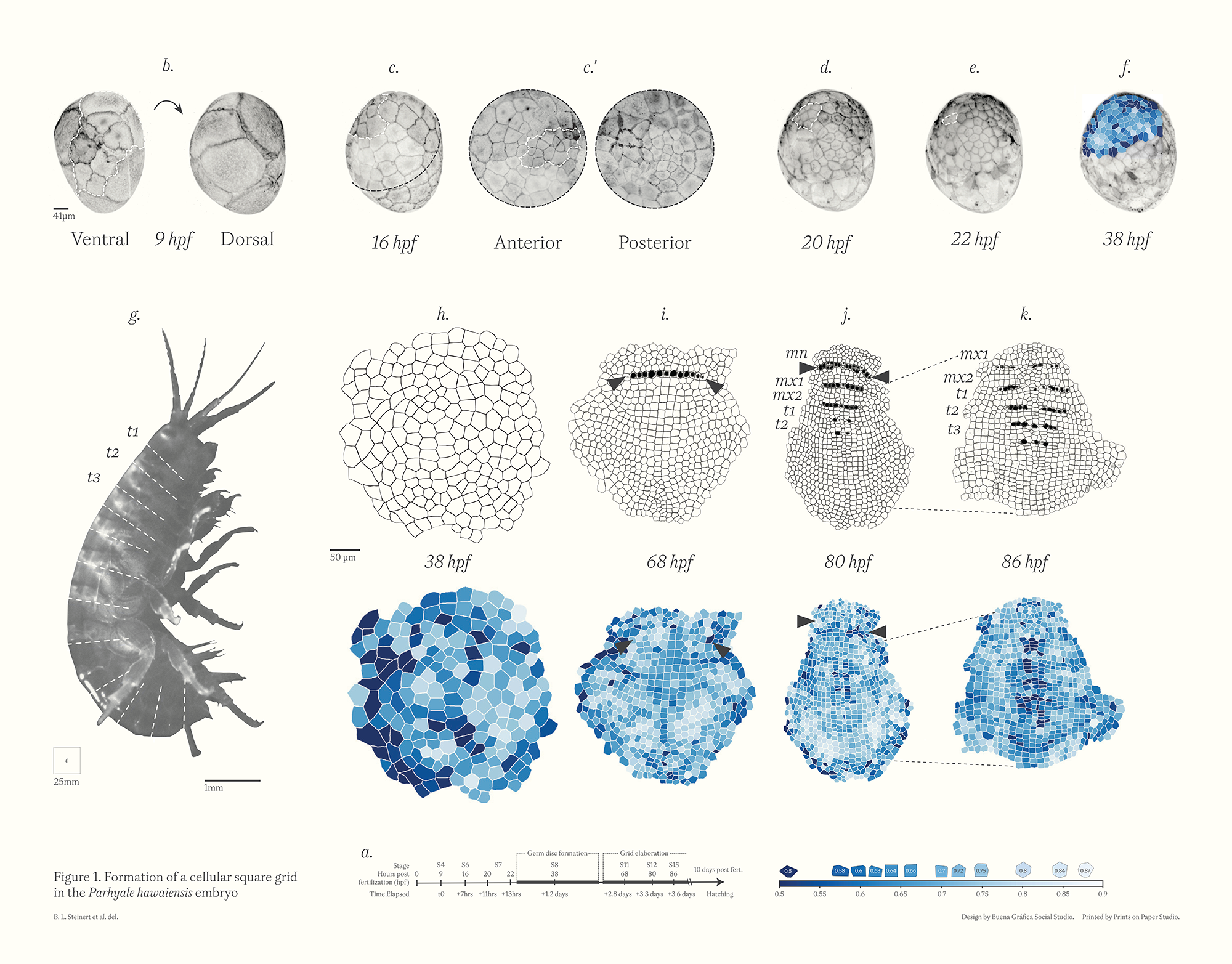
Figure 1 of “Cell lineage domains and cytoskeletal cables organize a cellular square grid in a crustacean,” redesigned as 36” x 48” 19th-century scientific wall chart. Designed in collaboration with Buena Gráfica Social Studio.
The prints and wall chart produced for Figure 1. In the Beginning were mounted as an exhibit in the lobby of the Harvard Science Center in 2025.
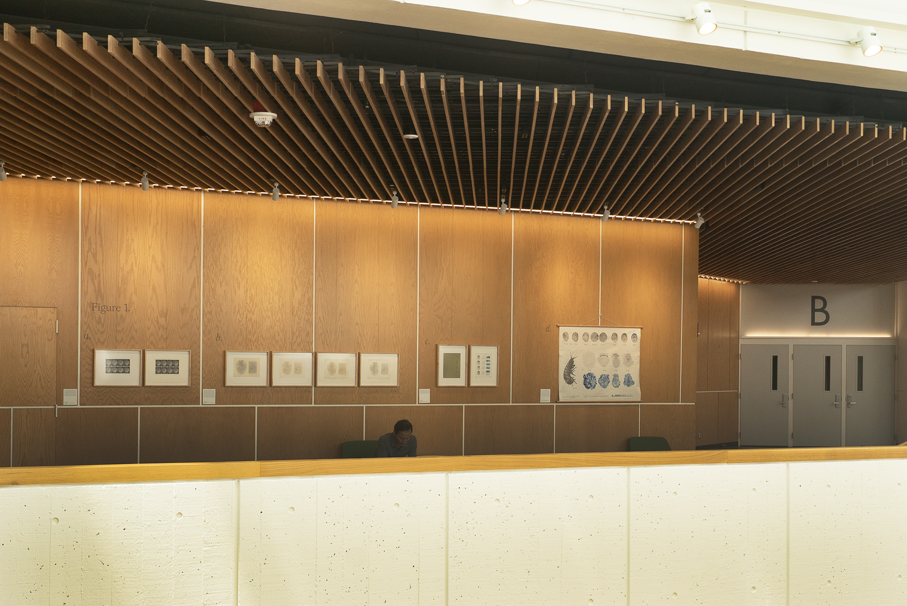
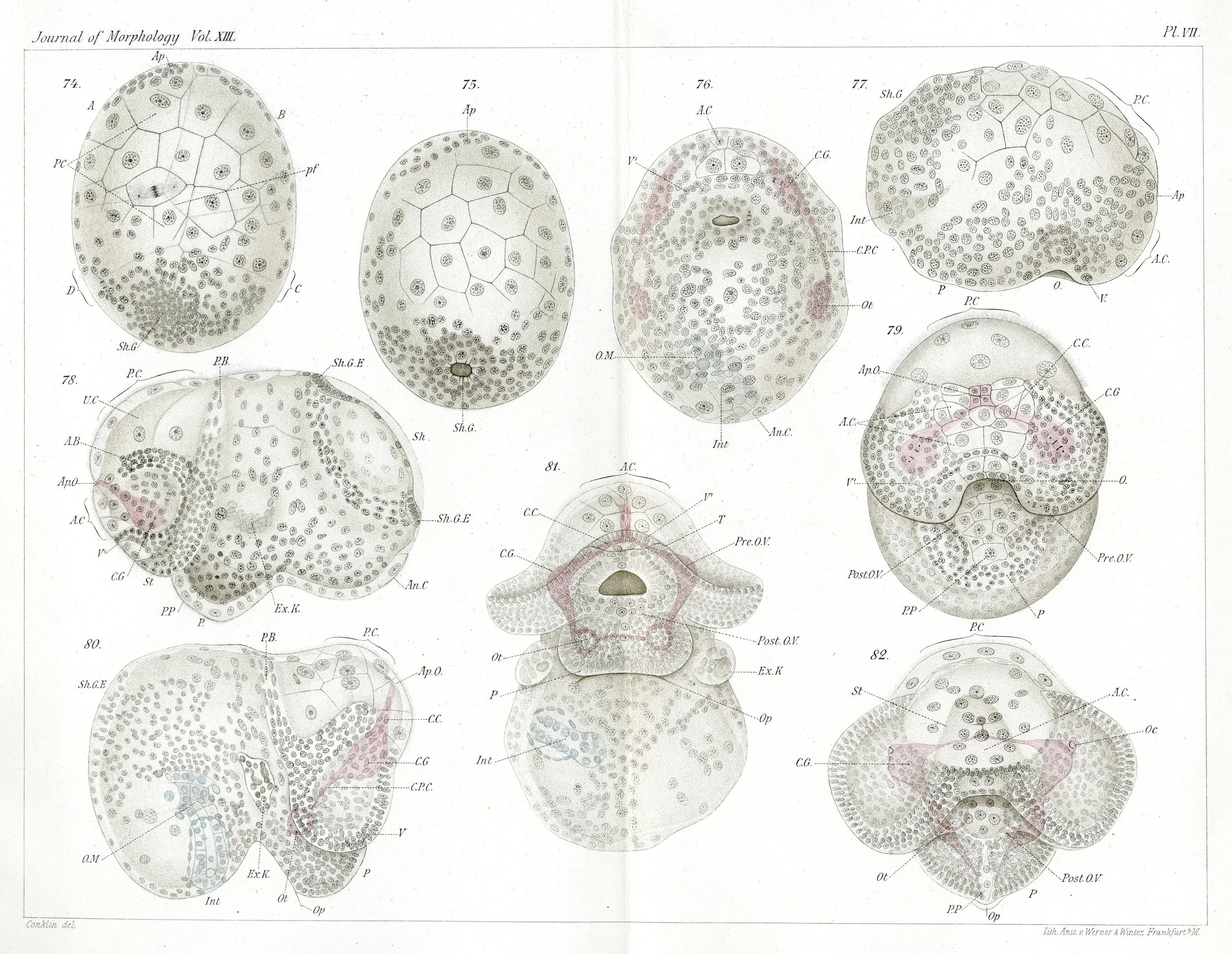
Figure 1 installed in the Harvard Science Center (left) and a lithographed fold-out figure from E.G. Conklin’s paper “The Embryology of Crepidula,” published in 1897 (right).
Design for a photogravure plate (left) printed (right) at Prints on Paper Studio in Cabot, VT. 11” x 14” inches on Hahnemühle paper. The figure panels in this plate are drawn from my paper “Cell lineage domains and cytoskeletal cables organize a cellular square grid in a crustacean.”