Morphogenesis
Nuclei in a Parhyale hawaiensis embryo germ band.
Most of the organisms we encounter on a daily basis emerged from a single cell — an egg, seed, or spore — or small cluster of cells. My research explores morphogenesis, the developmental process through which the forms of multicellular organisms emerge. I look at the dynamic mechanisms that arise from interactions among molecules, genes, cells, and structurally integrated tissues. I’m particularly interested in how the geometries and mechanics of cells carry information that regulates cell-fate determination as well as how the cytoskeleton (actin, non-muscle myosin, and microtubules) physically sculpts the shapes of organisms. I primarily work with marine invertebrates due to the relative transparency and beauty of their embryos, and the range of developmental strategies they have evolved. My work is centered around long-term live-imaging with confocal and lightsheet microscopy and mechanical and molecular perturbations to understand morphogenetic mechanisms.
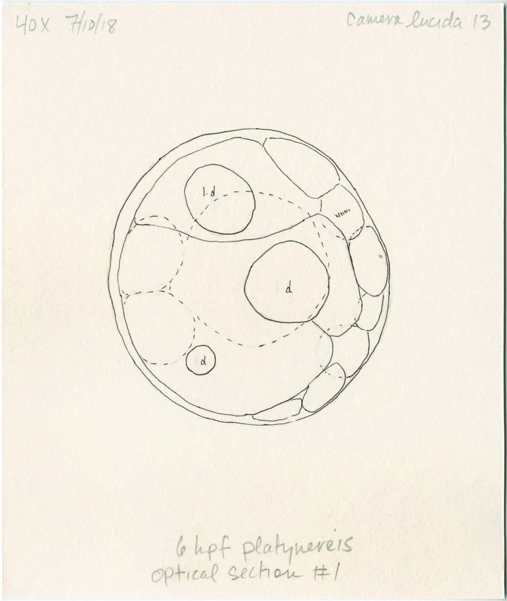
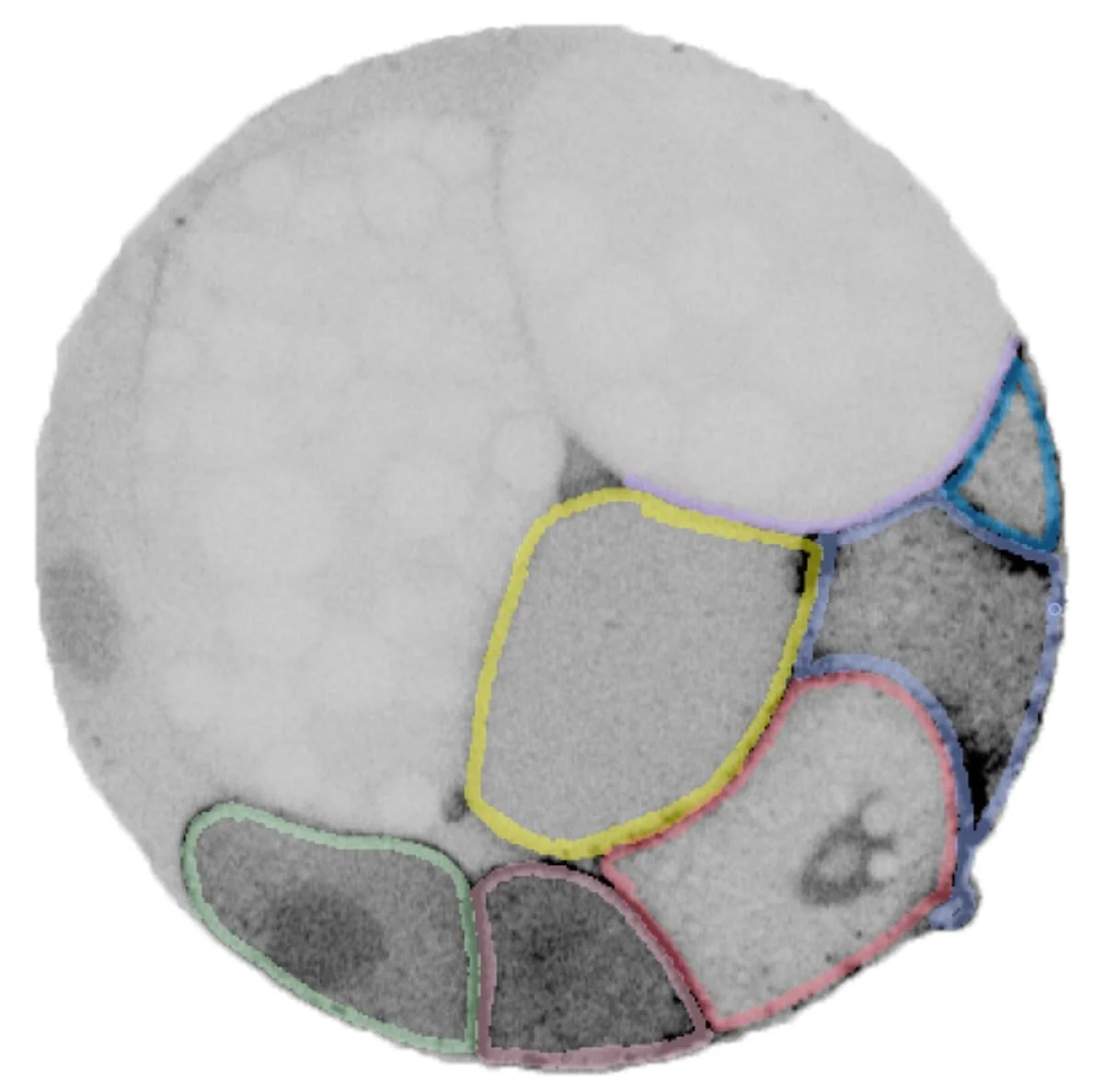
32-cell Platynereis dumerilii embryo. Camera lucida drawing (left), segmented cells in a confocal microscope z-section (middle), and full embryo 3D model (right). The central micrograph 3D model are of the same specimen.
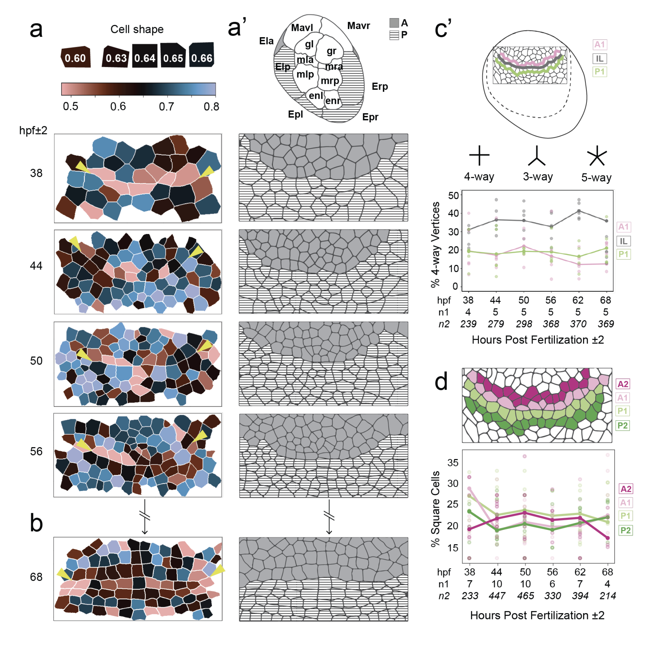
Figures 2 (left) and 4 (right) from my paper “Lineage domains and cytoskeletal cables organize a cellular square grid in a crustacean,” depicting the emergence of body-plan organization and formation of square cells in this embryo.
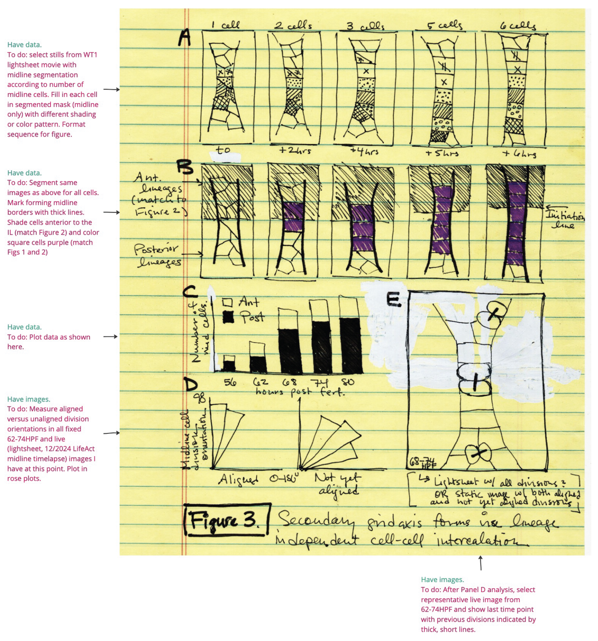
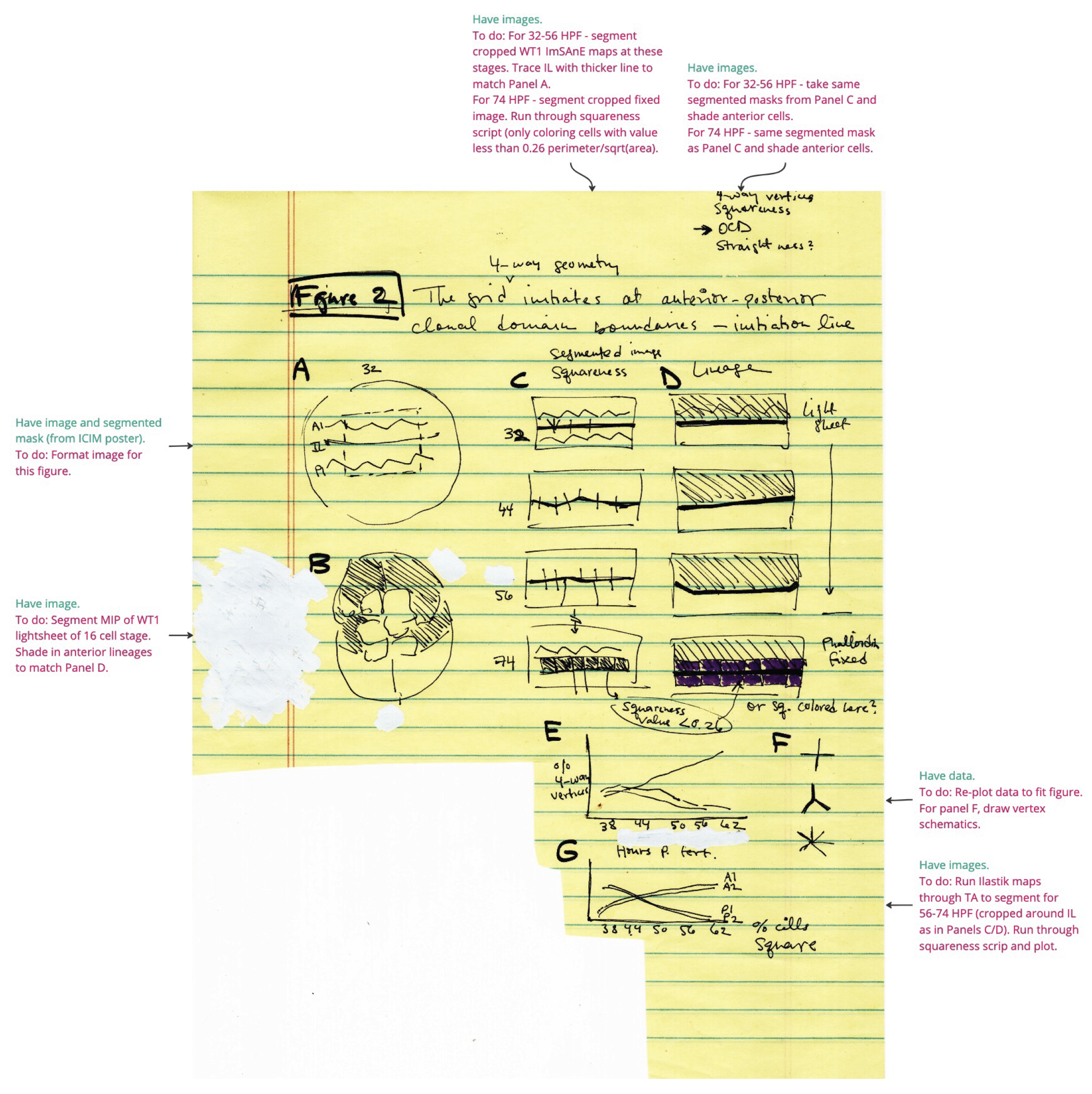
Sketches and experimental plans for Figures 2 (left) and 4 (right, formerly Figure 3) from “Lineage domains and cytoskeletal cables organize a cellular square grid in a crustacean.”
I am fascinated by how what we can know about the natural world is inseparable from how we see it. At the microscopic scale, we have no other way to know organisms other than through lens-based instrumentation, significant manipulation of the tissue, and some form of image-making. A confocal time-lapse sequence of a florescent-labeled tissue, an animation of a 3D rendering, or visualization of single-cell RNA sequencing for example, each provides a different view of life at this scale.
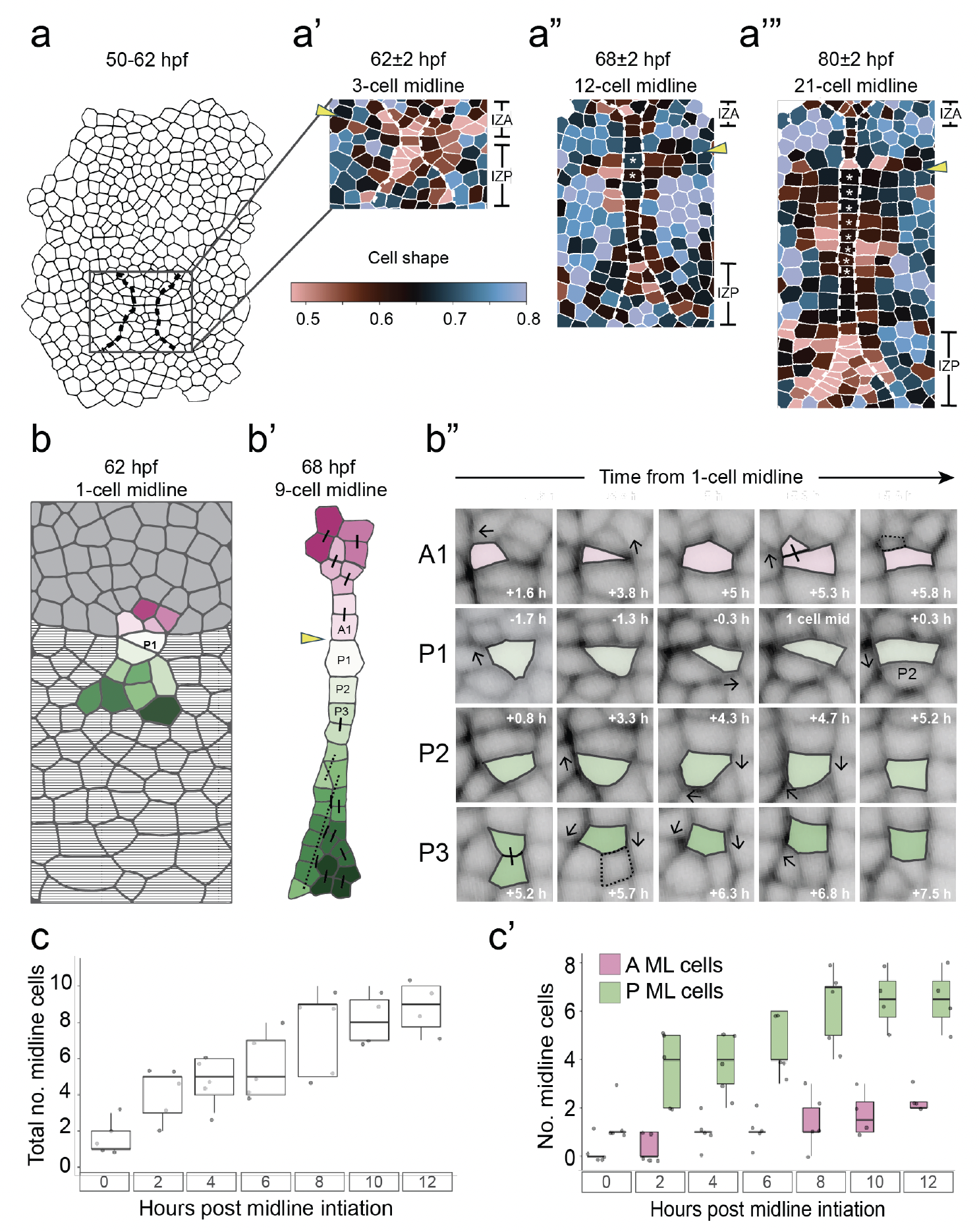
Figures 2 (left) and 4 (right) from my paper “Lineage domains and cytoskeletal cables organize a cellular square grid in a crustacean,” depicting the emergence of body-plan organization and formation of square cells in this embryo.
Much of biology involves grappling with the nuances of change over time and through space. I love thinking about how to capture, comprehend, and represent the movement of cells or sub-cellular structures with microscopes and various image-analysis modalities. I see visual communication and scientific figure design as fundamental to this work. Clear visual storytelling is essential not only for effectively sharing knowledge about the living world but also for how we as researchers understand it ourselves.
I begin sketching figures early in the research process as a way of thinking through what kind of story I want to tell, planning experiments, and filling in data analysis gaps.

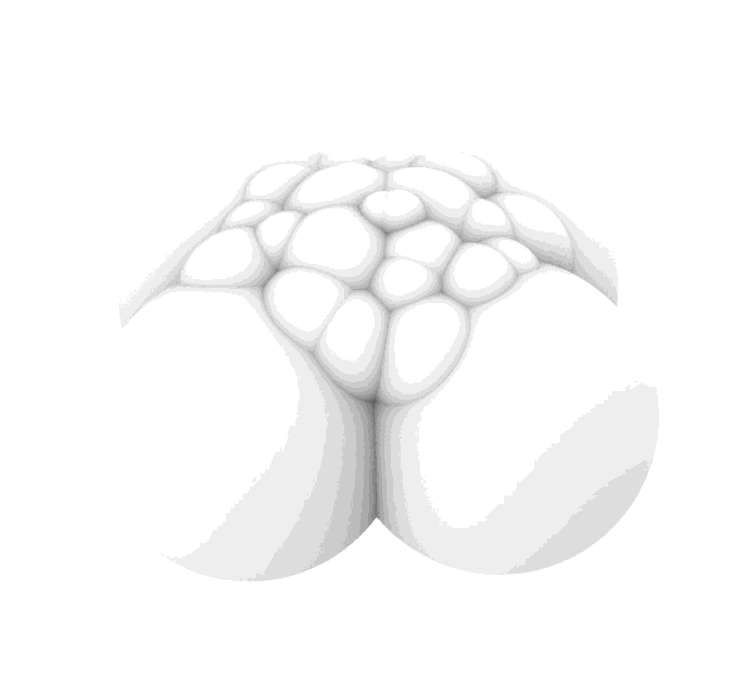
Development of the Parhyale hawaiensis germ band, the embryonic structure that establishes the blueprint for the adult body. On the left is a 16-cell stage embryo and on the right is the same embryo approximately two days later. This time-lapse was generated on a multiview lightsheet microscope.
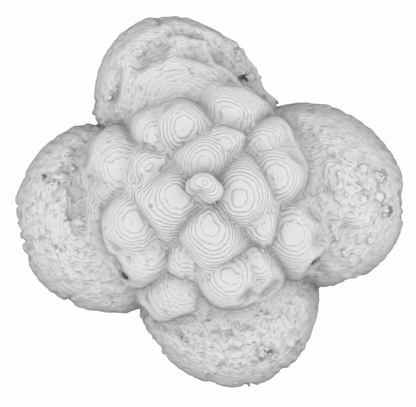
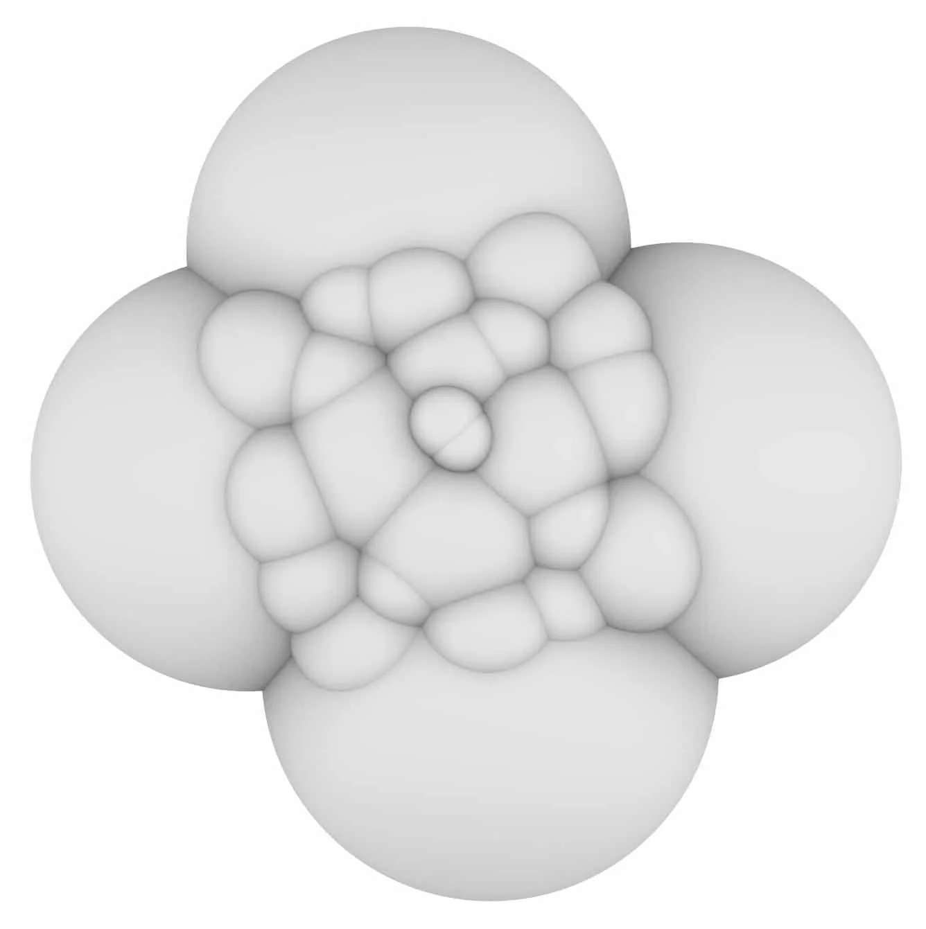
3D model of a 24-cell Crepidula fornicata embryo. Raw data from a confocal z-stack (left) and extrapolated model (center and right) generated in collaboration with Matt Muller at Pneuhaus.
I have found that while advanced image analysis, including machine-learning based modalities, now allows us to see these processes in novel and exciting ways, it is also in many ways upending the embodied observational practices that have been central to biology for centuries. To address this, I am exploring ways of using virtual reality to engage with microscope image datasets.
Visualizing Crepidula fornicata embryos in VR and under the microscope. Produced by Hyacinth Empinado and published in STAT News (2017) and Scientific American (2017)